The Cell's Transformations: Shrink, Grow, Switch, Die
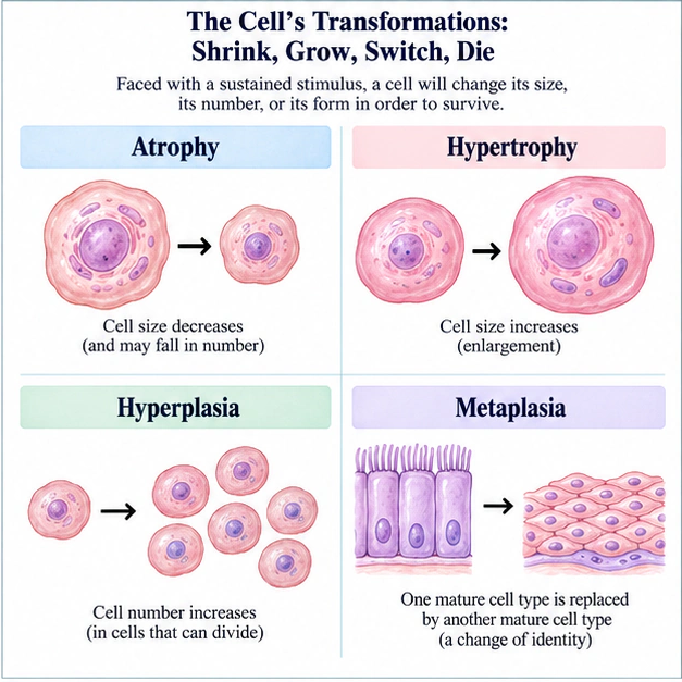
Metaplasia = a change of identity to adapt; hyperplasia = make more cells; hypertrophy = bulk up the cell; atrophy = smaller and fewer.
Focus points for this chapter
Scan the topics first, then read the chapter in order.
- Atrophy Is Not Simply "Getting Smaller"
- Metaplasia: Switching Epithelium to Suit the Environment
- Intracellular "Stockpiles": Before Leaping from What You See to a Name, Ask Why
- Reversible vs Irreversible: The Line Is Drawn at the Nucleus
- The Ceiling on Regeneration: Who Can Be Replaced, and Who Never Returns
Study illustration: The Cell's Transformations: Shrink, Grow, Switch, Die
Full text
In the pathology practical, a resident faces three slides and gives one answer for each: "Disuse muscle atrophy — the cells merely shrink; their number stays the same." "Barrett esophagus — that is hyperplasia." "After recurrent bladder infections, transitional epithelium replaced by transitional epithelium — that is metaplasia." The attending smiles and shakes his head: three answers, all three wrong. Where is the error? In never first asking "why is the cell doing this?"
Faced with a sustained stimulus, a cell will "change its size, its number, or its form" in order to survive. At first glance the questions seem to test a lookup table matching atrophy, hypertrophy, hyperplasia, and metaplasia to examples; the real key is to think through what the stimulus is, whether the cell is capable of dividing, and whether it ends up changing its dimensions or its identity. Once you grasp this logic, the table grows in your mind by itself.
Atrophy Is Not Simply "Getting Smaller"
Study illustration: Atrophy Is Not Simply "Getting Smaller"
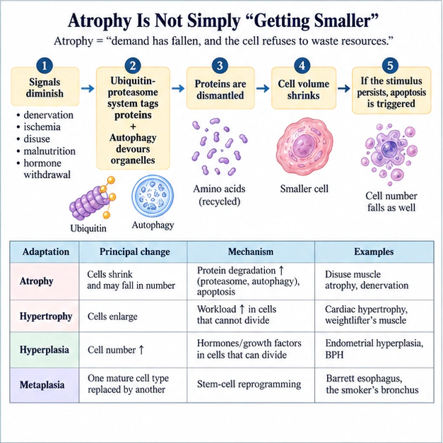
The true meaning of atrophy is "demand has fallen, and the cell refuses to waste resources," and behind it lies a five-step causal chain: ① signals diminish (denervation, ischemia, disuse, malnutrition, hormone withdrawal) → ② the ubiquitin-proteasome system tags proteins + autophagy devours organelles → ③ proteins are dismantled → ④ cell volume shrinks → ⑤ if the stimulus persists, some cells trigger apoptosis and exit → cell number falls as well. So the statement "atrophy = only smaller, number unchanged" drops the fifth step — in long-standing atrophy the cells become both smaller and fewer.
- Correct answers: atrophy = smaller and fewer; hypertrophy = bulk up; hyperplasia = make more; metaplasia = change identity.
- Trap one: "atrophy only shrinks cells, number unchanged" — omits the apoptosis step; wrong.
- Trap two: BPH may be spelled out as "benign prostatic hypertrophy," but in essence it is hyperplasia, not hypertrophy.
- Trap three: classifying an enlarging myocardium as "hyperplasia" — cardiac myocytes do not divide; they can only hypertrophy.
Full text · 1 table
Hypertrophy is the exact opposite: when a cell's capacity to divide is limited (cardiac muscle, skeletal muscle) yet it is loaded with extra work for a long time, it cannot "make more of itself" and can only "bulk itself up" — this is hypertrophy, driven by increased protein synthesis. Cells that can divide, when stimulated by growth factors or hormones, undergo "hyperplasia" — an increase in number, as in endometrial hyperplasia or prostatic enlargement (BPH is in fact hyperplasia, not true hypertrophy). Put simply: cells that cannot divide enlarge themselves (hypertrophy); cells that can divide make more of themselves (hyperplasia).
| Adaptation | Principal change | Mechanism | Examples |
|---|---|---|---|
| Atrophy | Cells shrink and may fall in number | Protein degradation↑ (proteasome, autophagy), apoptosis | Disuse muscle atrophy, denervation |
| Hypertrophy | Cells enlarge | Workload↑ in cells that cannot divide | Cardiac hypertrophy, weightlifter's muscle |
| Hyperplasia | Cell number↑ | Hormones/growth factors in cells that can divide | Endometrial hyperplasia, BPH |
| Metaplasia | One mature cell type replaced by another mature cell type | Stem-cell reprogramming | Barrett esophagus, the smoker's bronchus |
Swipe or scroll sideways to compare every column; keyboard: focus the table and use arrow keys.
Metaplasia: Switching Epithelium to Suit the Environment
Study illustration: Metaplasia: Switching Epithelium to Suit the Environment
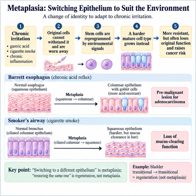
The core of metaplasia is "a change of identity," and behind it too runs a causal chain: ① chronic irritation (gastric acid, cigarette smoke, chronic inflammation) → ② the original, delicate cells cannot withstand it and are worn away → ③ the tissue's stem cells are reprogrammed by environmental signals → ④ they grow a hardier mature cell type instead → ⑤ hardier though it is, it often loses the original function and raises the risk of malignant change. Gastroesophageal reflux splashes acid onto the distal esophagus year after year; the squamous epithelium cannot hold, and the stem cells grow columnar epithelium with goblet cells instead — this is Barrett esophagus, more acid-resistant, but at the price of a higher cancer risk, making it a pre-malignant lesion for adenocarcinoma. Smoking sends heat and toxins into the airway; ciliated columnar epithelium is replaced by squamous epithelium, hardier yet stripped of its mucus-clearing function.
- Barrett esophagus = squamous→columnar (with goblet cells) = metaplasia = pre-malignant lesion for adenocarcinoma.
- The smoker's airway = ciliated columnar→squamous (hardier, but mucus clearance is lost).
- Trap one: bladder "transitional→transitional" is regeneration, not metaplasia (the same type does not count).
- Trap two: metaplasia = a reversible change of identity; dysplasia is where the pre-cancerous process truly begins.
- Trap three: calling Barrett "hyperplasia" is an error that simply gives the mark away.
Full text
Metaplasia has one criterion that is easily switched under your nose: the two cell types must be different. In recurrent bladder infection, urothelium sloughs and urothelium grows back; that is regeneration and hyperplasia, not metaplasia — replacement by the same cell type does not count. Examiners love this as a trap: the stem reads like metaplasia while the cell types are quietly tampered with. One sentence breaks it: "switching to a different epithelium" is metaplasia; "restoring the same one" is merely regeneration.
Intracellular "Stockpiles": Before Leaping from What You See to a Name, Ask Why
Study illustration: Intracellular "Stockpiles": Before Leaping from What You See to a Name, Ask Why
Full text · 1 table
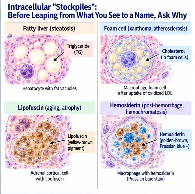
A liver section, its hepatocytes crammed with clear white vacuoles; beside it a skin section, the dermis filled with a cluster of cells whose cytoplasm looks like foam; beside that an adrenal gland, scattered with small yellow-brown nodules. Three kinds of "stockpile" — at first glance all just "something inside the cell" — yet what is stored is entirely different.
In questions on intracellular accumulations, the cardinal sin is "see foam, recite cholesterol." What you must remember is why the material is stored here: when hepatocytes are overloaded with fatty acids (alcohol, obesity, hypoxia), triglyceride (TG) is packaged into the cytoplasm as large vacuoles — this is steatosis; when macrophages in the arterial intima engulf oxidized LDL, their cytoplasm fills with cholesterol crystals and looks foamy — these are foam cells, the hallmark of atheromatous plaque and xanthoma; the yellow-brown lipofuscin in long-lived organs (heart, liver, adrenal) is a pigment accumulated over years of cellular metabolism, a badge of aging and wear-and-tear; and hemosiderin is the golden-brown pigment left after red cells are phagocytosed and metabolized following hemorrhage, positive on Prussian blue stain, seen after bleeding and in hemochromatosis.
| Accumulated material | Cellular appearance | Lesion |
|---|---|---|
| Triglyceride (TG) | Intracellular fat vacuoles | Steatosis (fatty liver, alcohol, hypoxia) |
| Cholesterol | Foam cells | Xanthoma, atherosclerosis |
| Lipofuscin | Yellow-brown pigment | Aging, atrophy (wear-and-tear) |
| Hemosiderin | Golden-brown, Prussian blue (+) | Post-hemorrhage, hemochromatosis |
Swipe or scroll sideways to compare every column; keyboard: focus the table and use arrow keys.
Reversible vs Irreversible: The Line Is Drawn at the Nucleus
Study illustration: Reversible vs Irreversible: The Line Is Drawn at the Nucleus
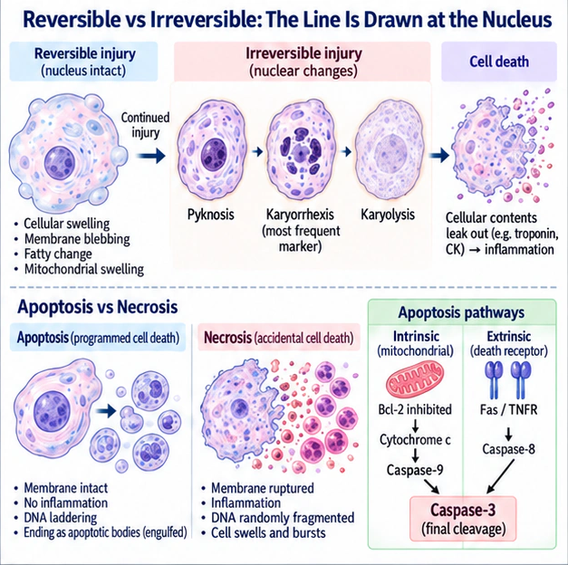
An injured cell can slide all the way to death, but the watershed between reversible and irreversible is written on the nucleus, in a five-step argument: ① oxidative phosphorylation falls → ATP is depleted → ② the sodium-potassium pump fails: cellular swelling, membrane blebbing, fatty change, mitochondrial swelling — so far the nucleus is intact and everything is still reversible → ③ once the mitochondrial permeability transition pore (MPTP) opens permanently and cytochrome c is released, the point of no return is crossed → ④ the nucleus proceeds through pyknosis → karyorrhexis → karyolysis → ⑤ the cell membrane ruptures and enzymes leak out (troponin and CK from myocardium, for instance), igniting inflammation. Of these, karyorrhexis is the most frequently named marker of irreversibility.
- Reversible = cellular swelling, nucleus intact; irreversible = pyknosis → karyorrhexis → karyolysis (karyorrhexis is the marker).
- Apoptosis: membrane intact, no inflammation, DNA laddered; necrosis: membrane ruptured, inflammation, DNA randomly fragmented.
- Intrinsic = cytochrome c → caspase-9; extrinsic = Fas/TNFR → caspase-8; both end at caspase-3.
- Trap one: treating "cellular swelling" as irreversible — it is still in the reversible stage.
- Trap two: assigning caspase-8 to the mitochondrial pathway — backwards.
- Trap three: does apoptosis provoke inflammation? The apoptotic membrane stays intact — it does not.
Full text
The contrast between apoptosis and necrosis is another line of easy marks. Apoptosis is programmed suicide (programmed cell death): active, energy-consuming, a single cell quietly dismantling itself, membrane intact, no inflammation, ending as apoptotic bodies that are engulfed; necrosis is being struck dead by accident: passive, the cell swells and bursts, enzymes leak, inflammation floods in. The two initiating pathways of apoptosis are also tested, and behind them lies a complete enzymatic cascade: the intrinsic pathway runs through the mitochondrion — Bcl-2 is inhibited, cytochrome c is released, caspase-9 is activated; the extrinsic pathway runs through death receptors — Fas/TNFR binding activates caspase-8. Both converge on caspase-3, which carries out the final cleavage and yields DNA laddering (fragments in multiples of 180 bp) — utterly unlike necrosis, where DNA is chopped into random sizes; this is the discriminating detail in high-resolution questions. Collect the three major patterns of necrosis while you are here: coagulative (ischemia of heart and kidney), liquefactive (brain, suppuration), caseous (TB).
The Ceiling on Regeneration: Who Can Be Replaced, and Who Never Returns
Study illustration: The Ceiling on Regeneration: Who Can Be Replaced, and Who Never Returns
- Steatosis = TG; xanthoma foam cells = cholesterol; lipofuscin = aging; hemosiderin = hemorrhage/iron deposition, Prussian blue positive.
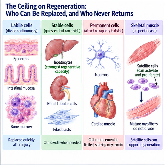
- Strongest regenerative capacity = liver (a stable cell); neurons and cardiac muscle have very limited regenerative capacity; mature skeletal muscle fibers do not divide, but satellite cells can support repair.
- Trap one: fatty liver stores cholesterol? Wrong — it is TG.
- Trap two: foam cells store TG? Wrong — it is cholesterol.
- Trap three: the liver is made of labile cells? Wrong — it is the stable cell with the strongest regenerative power.
Full text
A tissue's capacity to regenerate is decided by the proliferative class of its cells. Labile cells — epidermis, intestinal mucosa, bone marrow — divide continuously and are replaced quickly after injury. Stable cells — hepatocytes, renal tubular cells, fibroblasts — are quiescent at rest yet can divide when needed; among them the hepatocyte is ranked as having the strongest regenerative capacity, which is why the liver refills rapidly after partial hepatectomy and why living-donor liver transplantation is feasible at all. Permanent cells — neurons and cardiac muscle — have almost no capacity to divide, and after injury their function can only be patched with scar; this explains why a myocardial infarction leaves fibrous scar rather than new myocardium, and why nerve injury so often fails to recover completely. Skeletal muscle requires a distinction: mature myofibers do not divide, but satellite cells can activate, proliferate and participate in regeneration. Skeletal muscle tissue is therefore not wholly incapable of repair.
Smaller and fewer? Atrophy!
Heart bulks up? Hypertrophy!
B-P-H makes more? Hyperplasia!
Barrett goes columnar? Metaplasia!
Fatty liver T-G, foam cell cholesterol
Caspase nine intrinsic, eight extrinsic, three's the call
🎤 English rap lyrics — the chapter's study song (chorus = the mnemonics above)
Shrink, Grow, Switch, Die
Verse 1
Atrophy: demand falls, it won't waste resources,
Denervation, disuse, ischemia are the sources,
Ubiquitin-proteasome, autophagy: it shrinks,
Stress drags on, apoptosis kicks in, the count sinks,
Can't divide, heart, skeletal? Load it, cells enlarge,
Can divide, hormones, growth factors? Numbers take charge,
Big heart's never hyperplasia, myocytes don't split,
B-P-H spells "hypertrophy," but hyperplasia's it,
Chronic irritation? Stem cells switch the design,
Hardier, but function's lost and cancer risk climbs,
Reflux burns squamous, columnar with goblets, new persona,
Barrett's pre-malignant for adenocarcinoma,
Smoke turns ciliated columnar squamous, mucus sweep is done,
Metaplasia can reverse; dysplasia's where pre-cancer's begun,
Transitional to transitional? Type stays the same,
That's regeneration, metaplasia's the wrong name.
Chorus
Smaller and fewer? Atrophy!
Heart bulks up? Hypertrophy!
B-P-H makes more? Hyperplasia!
Barrett goes columnar? Metaplasia!
Fatty liver T-G, foam cell cholesterol
Caspase nine intrinsic, eight extrinsic, three's the call
Verse 2
Liver stuffed with T-G vacuoles? Steatosis,
Xanthoma foam cells? Cholesterol, atherosclerosis,
Hemosiderin after bleeding stains Prussian blue,
Lipofuscin, yellow-brown, aging's wear-and-tear residue,
Sodium-potassium pump fails when A-T-P's low,
Swelling looks bad, nucleus intact: reversible though,
M-P-T-P opens for good, cytochrome c goes free,
Pyknosis, karyorrhexis, karyolysis, one-two-three,
Karyorrhexis: the marker named for no return,
Membrane ruptures, enzymes leak, inflammation burns,
Apoptosis spends energy, membrane intact, no inflammation's spread,
D-N-A ladders, multiples of one-eighty; necrosis, random instead.
Chorus
Smaller and fewer? Atrophy!
Heart bulks up? Hypertrophy!
B-P-H makes more? Hyperplasia!
Barrett goes columnar? Metaplasia!
Fatty liver T-G, foam cell cholesterol
Caspase nine intrinsic, eight extrinsic, three's the call
Bridge
Intrinsic: B-C-L-two blocked, cytochrome c, caspase-nine,
Extrinsic: Fas, T-N-F-R, caspase-eight in line,
Coagulative: ischemic heart, kidney. Caseous: T-B,
Liquefactive: brain, suppuration, that's the three,
Liver: stable, not labile, regrowth's number-one star,
Neurons, heart, skeletal muscle? Permanent, patched with scar.
Chorus
Smaller and fewer? Atrophy!
Heart bulks up? Hypertrophy!
B-P-H makes more? Hyperplasia!
Barrett goes columnar? Metaplasia!
Fatty liver T-G, foam cell cholesterol
Caspase nine intrinsic, eight extrinsic, three's the call
Outro
All that swelling's reversible, nucleus intact,
Is it karyorrhexis? No way back,
On regrowth the liver leads; neurons, heart just scar,
Lucid: apoptosis, membrane intact, no inflammation.
Close the book and recall
Say the answer in your own words or jot down keywords, then check each one. This is recall practice, not a past exam question.
What can you recall about “The Four Adaptations · Traps”?
Once you have an answer, open the original to check
★ Must-know text from this chapter
The Four Adaptations · Traps
- Correct answers: atrophy = smaller and fewer; hypertrophy = bulk up; hyperplasia = make more; metaplasia = change identity.
- Trap one: "atrophy only shrinks cells, number unchanged" — omits the apoptosis step; wrong.
- Trap two: BPH may be spelled out as "benign prostatic hypertrophy," but in essence it is hyperplasia, not hypertrophy.
- Trap three: classifying an enlarging myocardium as "hyperplasia" — cardiac myocytes do not divide; they can only hypertrophy.
What can you recall about “Metaplasia · Traps”?
Once you have an answer, open the original to check
★ Must-know text from this chapter
Metaplasia · Traps
- Barrett esophagus = squamous→columnar (with goblet cells) = metaplasia = pre-malignant lesion for adenocarcinoma.
- The smoker's airway = ciliated columnar→squamous (hardier, but mucus clearance is lost).
- Trap one: bladder "transitional→transitional" is regeneration, not metaplasia (the same type does not count).
- Trap two: metaplasia = a reversible change of identity; dysplasia is where the pre-cancerous process truly begins.
- Trap three: calling Barrett "hyperplasia" is an error that simply gives the mark away.
What can you recall about “Accumulations · Regeneration · Traps”?
Once you have an answer, open the original to check
★ Must-know text from this chapter
Accumulations · Regeneration · Traps
- Steatosis = TG; xanthoma foam cells = cholesterol; lipofuscin = aging; hemosiderin = hemorrhage/iron deposition, Prussian blue positive.
- Strongest regenerative capacity = liver (a stable cell); neurons and cardiac muscle have very limited regenerative capacity; mature skeletal muscle fibers do not divide, but satellite cells can support repair.
- Trap one: fatty liver stores cholesterol? Wrong — it is TG.
- Trap two: foam cells store TG? Wrong — it is cholesterol.
- Trap three: the liver is made of labile cells? Wrong — it is the stable cell with the strongest regenerative power.
After checking, reread whatever you missed or mixed up, then try once more without looking.
★ High-yield points & traps from past exams (1 section)
| Exam point | Correct answer | Common trap |
|---|---|---|
| Material accumulating in xanthoma foam cells | Cholesterol (lipid) | Answering triglycerides |
| Correct statement about atrophy | Cells shrink and their number may decrease | Choosing "number unchanged" by mistake |
| Material accumulating in fatty change | Triglycerides | Answering cholesterol |
| Hallmark of irreversible injury | Karyorrhexis and other nuclear changes | Mistaking cell swelling for irreversible injury |
| Barrett esophagus | GERD → squamous→columnar metaplasia (precursor lesion of adenocarcinoma) | Calling it hyperplasia by mistake |
| Bladder transitional epithelium replaced by transitional epithelium | Not metaplasia (same cell type) | Misjudging it as metaplasia |
| Organ with the greatest regenerative capacity | Liver (hepatocytes, stable cells) | Answering nerve/cardiac muscle |
| Neurons, cardiac myocytes | Permanent cells, do not regenerate | Thinking they can regenerate |
| Apoptosis vs necrosis | Apoptosis does not cause inflammation, membrane intact; necrosis triggers inflammation, membrane ruptured | Treating apoptosis as inflammatory |
| Key to the intrinsic apoptotic pathway | Mitochondrial cytochrome c → caspase-9 (inhibited by Bcl-2) | Answering caspase-8 (that is the extrinsic pathway) |
Swipe or scroll sideways to compare every column; keyboard: focus the table and use arrow keys.